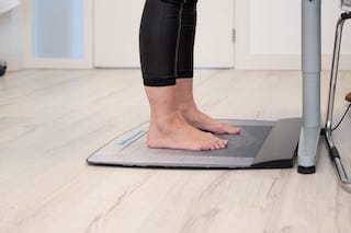
In de praktijk wordt gewerkt met apparatuur voor het digitaal analyseren van het looppatroon. Dit is een belangrijk onderdeel om de diagnose te stellen. Vaak is een visuele analyse onvoldoende. Dit drukmeetsysteem geeft door de vele ingebouwde sensoren informatie over de statische en dynamische belasting van de voet. Door bestudering van de gait-line en de drukverdeling tijdens het lopen, komen we meer te weten over het functioneren en de afwijkingen tijdens het lopen.
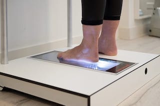
Voor het aanmeten van onze zolen wordt er tevens gebruik gemaakt van een 3D scanner. Het aangemeten voetbed wordt 1 op 1 verwerkt in de therapeutische zool waardoor deze perfect op uw voet aansluit.
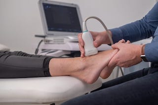
Het echografisch onderzoek wordt in onze praktijk uitgevoerd door gekwalificeerde en ervaren echografisten (Advanced practitioner MSU voet en enkel). Echografie is een laagdrempelig en pijnloos onderzoek. Het voordeel is dat er direct gekeken kan worden naar een eventuele aandoening. U weet dus snel of er iets aan de hand is.
U kunt direct een afspraak maken voor een echo, hiervoor heeft u geen verwijzing nodig.
Uiteraard kunnen wij desgewenst zorgen voor een adequaat verslag aan uw huisarts, fysiotherapeut of podotherapeut.
Echografie wordt toegepast om aanvullend beeldvormende informatie te krijgen over diverse structuren in de voet en enkel. Het kan worden ingezet voor het in beeld brengen van onder andere onderstaande structuren:
We zien o.a. veel patiënten met hielpijn, met klachten onder de voorvoet en met klachten in de voetwortel. Met een juiste diagnose kan er gerichter therapie worden ingezet. We werken intensief samen met fysiotherapeuten, orthopedische schoenmakers en orthopedisch specialisten bij vervolg trajecten. Onze echografist maakt deel uit van het team van het Hielpijncentrum.
De kosten voor een echografisch onderzoek zijn vanaf 70 euro en worden veelal vergoed vanuit uw aanvullende verzekering.